130
VOLUME 10 NUMBER 4 • NOVEMBER 2013
RESEARCH ARTICLE
SA JOURNAL OF DIABETES & VASCULAR DISEASE
DPOAE characteristics of both groups
An independent samples
t
-test indicated a statistically significant
difference in the scores for the left ear in the experimental and
control groups at 1 000 Hz (
p
= 0.04). No statistically significant
difference was found in the scores for both ears in the experimental
and control groups for the other frequencies (Table 3). The mean
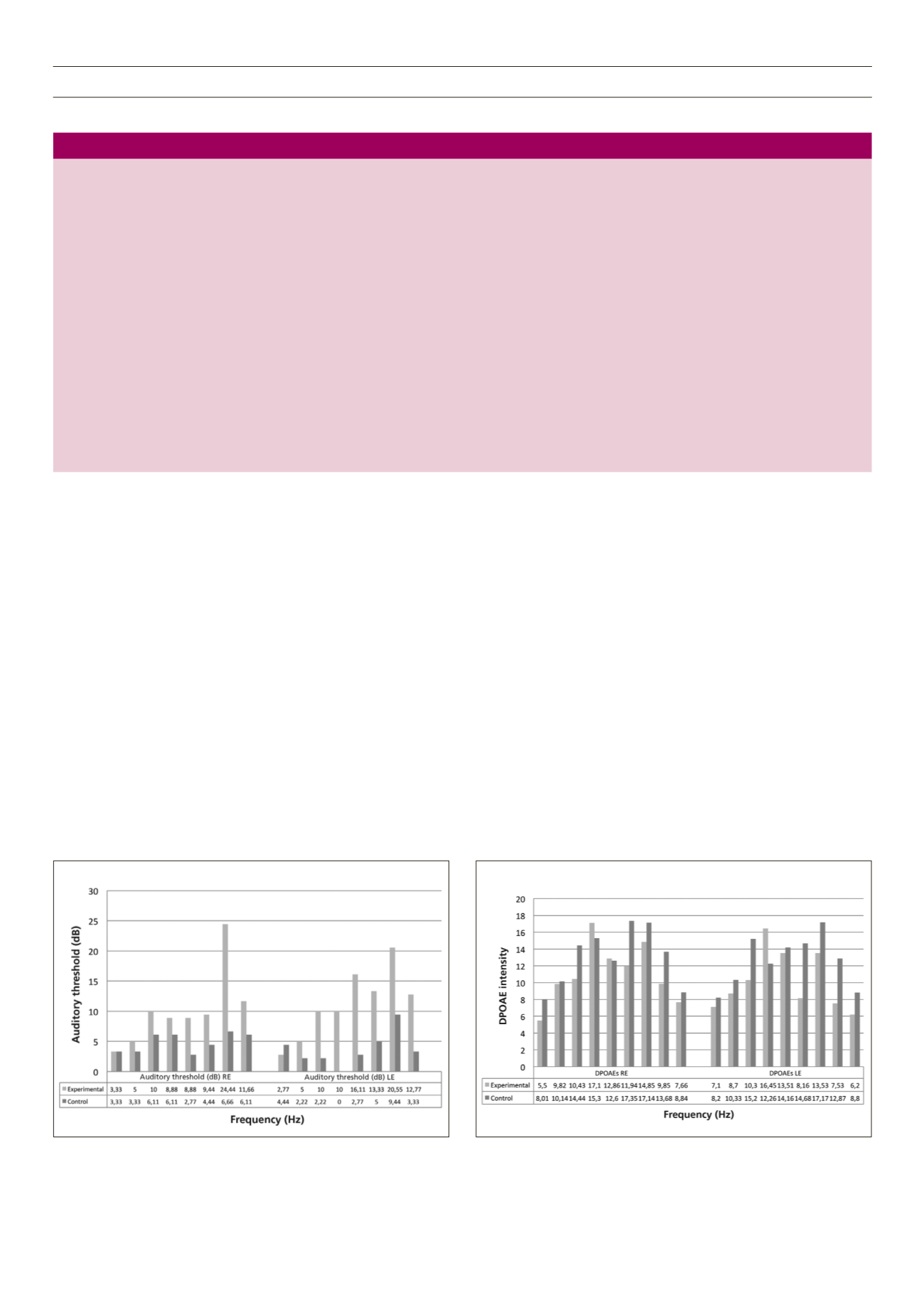
values of the right and left ear DPOAE characteristics indicate that
the DPOAE results were within normal limits for both groups at all
frequencies except 500 Hz in the experimental group (Fig. 2). The
reliability of the mean DPOAE amplitude at 500 Hz is questionable
as DPOAE amplitudes may fall below the lower end of the normal
range for all frequencies except 500 Hz.
27
The mean DPOAE values in the experimental group were
lower in the right and left ears than in the control group,
except at 1 500 Hz. This suggests that the mean DPOAE values
in the experimental group were closer to 6 dB, although within
normal limits, which may suggest an early sign of outer hair cell
(OHC) dysfunction. This is particularly important to note in the
current sample which comprised a relatively young age of T1DM
subjects.
DPOAE measures for detecting the early signs of cochlear
dysfunction in individuals with T1DM compared to pure-
tone test battery
The mean values of the right ear DPOAEs and pure tones (Fig. 3)
were within normal limits for all frequencies except at 500 Hz.
The auditory threshold value at 6 000 Hz, although within normal
limits, was the lowest compared to all other frequencies. The
mean values of the DPOAEs were found to be lower at 6 000 and
8 000 Hz compared to pure tones. This indicates that the DPOAE
measure may be more sensitive in detecting early signs of cochlear
dysfunction as the values are close to 6 dB.
The mean values of the left ear DPOAEs and pure tones
(Fig. 3) were within normal limits. The mean values of the DPOAEs
at 6 000 and 8 000Hz indicate that the DPOAE measure may be
more sensitive in detecting the early signs of cochlear dysfunction
Fig. 1.
Mean auditory threshold of left and right ears.
Fig. 2.
Mean DPOAE values of left and right ears.
Table 3.
p-
values of impedance audiometry, pure-tones audiometry and DPOAEs for left and right ears.
Tympanometry Right ear
Left ear
Static compliance 0.72
0.59
Ear canal volume
0.461
0.79
Ear pressure
0.4
0.96
Acoustic reflex
frequency (kHz)
0.5
1
2
4
0.5
1
2
4
Ipsilateral
0.8
0.9
0.3
0.6
0.8
0.5
0.7
0.6
Contralateral
0.15
0.43
0.8
0.71
0.43
0.52
0.91
0.87
Pure-tone
frequency (kHZ)
0.25 0.5 1
2
3
4
6
8
0.25 0.5 1
2
3
4
6
8
1
0.4 0
0
0
0.4 0
0
0.5 0.1 0
0
0
0
0
0.2
DPOAEs
0.5 0.75 1
1.5 2
3
4
6
8
0.5 0.75 1
1.5 2
3
4
6
8
frequency (kHZ)
0.18 0.88 0.14 0.61 0.93 0.08 0.56 0.25 0.56 0.39 0.32 0.04 0.95 0.83 0.08 0.26 0.16 0.55


