
RESEARCH ARTICLE
SA JOURNAL OF DIABETES & VASCULAR DISEASE
66
VOLUME 15 NUMBER 2 • NOVEMBER 2018
Renal function, as assessed by estimated glomerular filtration
rate (eGFR) using the Cockcroft Gault formula, was reasonably
preserved among the three groups. It was highest in the control
group but not statistically significantly different.
The mean values of all lipid components were normal and
comparable, except for the low-density lipoprotein (LDL) cholesterol
level and atherogenic ratio, which showed a significant stepwise
increase from control to microalbuminric group (
p
= 0.0008 and
p
= 0.01, respectively). FBS was also significantly higher in the
diabetic groups compared to the controls (
p
= 0.001).
Table 2 shows the echocardiographic parameters of LV function
among the three groups. Mean values of EF and FS were normal
in the three groups, but FS showed a significant stepwise decrease
from control to microalbuminuric group (
p
= 0.0002).
Doppler echocardiographic parameters showed some degree
of LV diastolic dysfunction, which was more pronounced in the
diabetic groups. A velocity (
p
= 0.0034), IVRT (
p
= 0.0001) and
PASP (
p
= 0.02) showed a significant stepwise increase from control
to microalbuminuric group, with a reverse trend for E velocity (
p
<
0.001) and E/A ratio (
p
< 0.001).
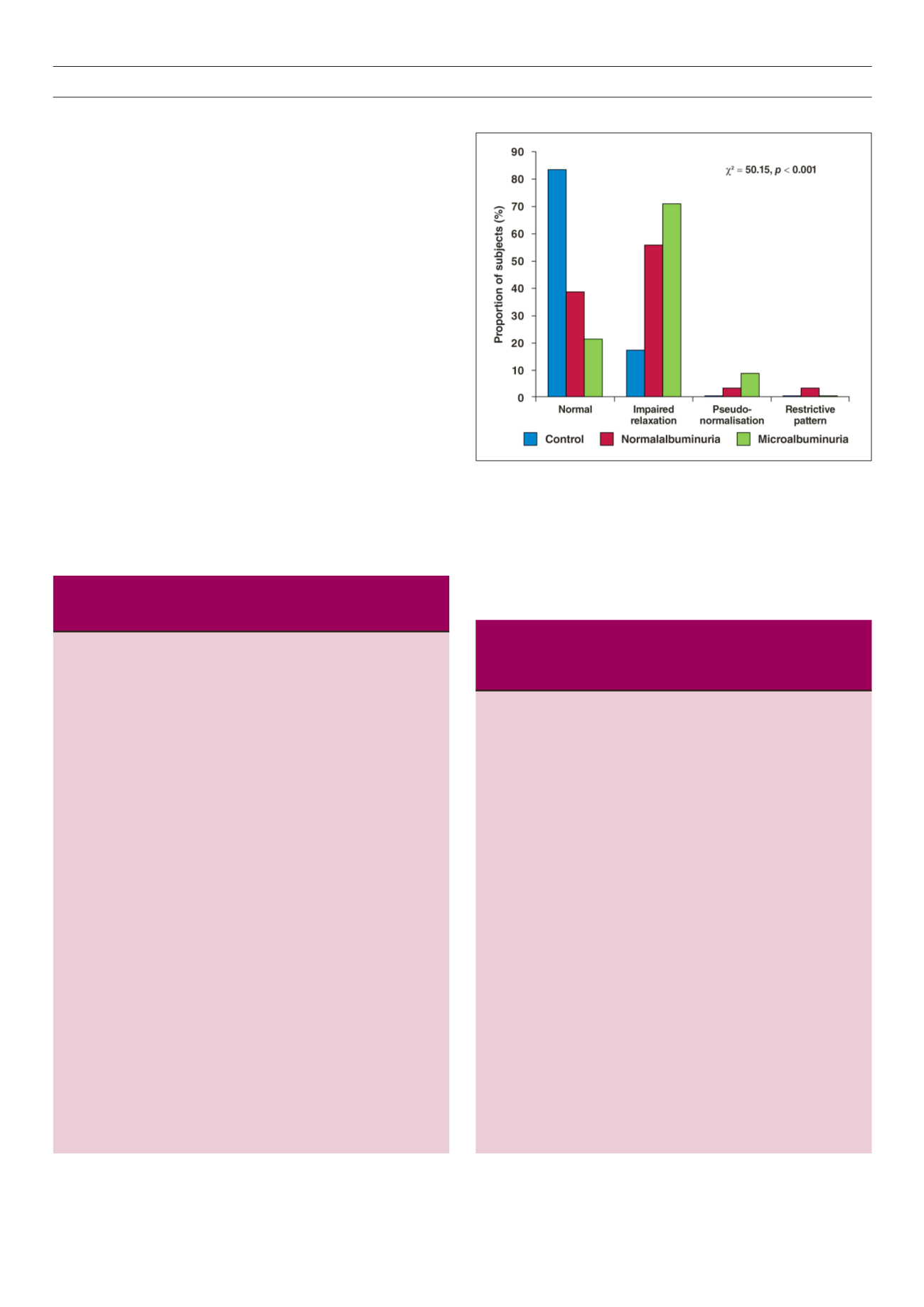
Fig. 1 shows the prevalence and pattern of LVDD among the
three groups. The prevalence of LVDD showed a stepwise increase
from 16.9% in the control to 78.9% in the microalbuminuric group.
The most common grade of DD was grade 1, which occurred in
70.4 and 55.5% of microalbuminuric and normoalbuminuric
groups, respectively, compared to 16.9% in the controls. Grade 1
was the only type of DD found in the control group; 3.2% of the
normoalbuminuric group and 8.5% of the microalbuminuric group
Table 2.
Comparison of echocardiographic parameters (mean ± SD) of
left ventricular systolic and diastolic function among healthy controls,
and normotensive diabetics with normoalbuminuria or micro-
albuminuria
Normo-
Micro-
Control albuminuric albuminuric
Echo parameters (
n
= 59) (
n
= 63)
(
n
= 71) F-test
p
-value
LVIDd (mm)
42 ± 4.4
†
40 ± 4.9
38 ± 4.3
7.84 .0006
LVIDs (mm)
27 ± 3.2 26 ± 2.8
26 ± 3.2
0.81 0.45
EDV (ml)
82 ± 19
†
76 ± 20
68 ± 16
8.13 0.0004
ESV (ml)
27 ± 8
26 ± 8
25 ± 7
0.82 0.44
Stroke volume (ml) 54 ± 20
†
51 ± 19
†
45 ± 17
9.28 0.0002
Cardiac output (l)
4.3 ± 0.9
†
4.2 ± 1.1
†
3.6 ± 1.0 10.05 0.0002
Ejection fraction (%) 62 ± 7.3
63 ± 8
60 ± 6.2
1.99 0.14
FS (%)
36 ± 5.5
†
34 ± 6.1
†
31 ± 4.1 11.39 < 0.001
Mitral E velocity (m/s) 77 ± 21*
†
65 ± 18
61 ± 12
20.65 < 0.001
Mitral A velocity (m/s) 67 ± 17
†
69 ± 13
74 ± 13
5.9 0.0034
E/A ratio
1.2 ± 0.3
†
1.0 ± 0.3
0.8 ± 0.2 31.51 < 0.001
IVRT (s)
79 ± 13
†
84 ± 16
90 ± 18
9.65 0.0001
Deceleration time (s) 199 ± 29 192 ± 42
192 ± 33
0.03 0.9658
PVF S velocity (m/s) 56 ± 11
†
47 ± 14
52 ± 11
4.57 0.0123
PVF D velocity (m/s) 49 ± 8*
42 ± 7
47 ± 11
5.11 0.0074
S/D ratio
1.2 ± 0.2 1.1 ± 0.2
1.1 ± 0.3
0.38 0.685
PVF Ar velocity (m/s) 31 ± 4.4 33 ± 4.0
34 ± 3.0
2.72 0.069
PASP (mmHg)
30 ± 8
30 ± 7
†
33 ± 9
4.22 0.0165
LADs (mm)
35 ± 3.3 34 ± 3.5
†
36 ± 3.6
4.5 0.0125
*
p
< 0.05 compared to normoalbuminuria by ANOVA followed by Bonferroni
post hoc
test.
†
p
< 0.05 compared to microalbuminuria by ANOVA followed by Bonferroni
post hoc
test.
F-test for ANOVA.
PVF: pulmonary venous flow, LADs: left atrial end-systolic dimension, IVRT:
isovolumic relaxation time, E: transmitral early-to-late inflow velocity ratio, A:
transmitral late atrial velocity, PASP: pulmonary artery systolic pressure.
Table 1.
Sociodemographic, anthropometric and laboratory data (mean
± SD) variations among healthy controls, and normotensive diabetics
with normoalbuminuria or microalbuminuria
Normo-
Micro-
Controls albuminuric albuminuric
Characteristics
(
n
= 59)
No = 63 (
n
= 71) F-test
p
-value
Age (years)
47 ± 10.0
50 ± 7.5
51 ± 7.0
0.87 0.43
Gender (% male)
49
51
45
2.05 0.36
DMdur (years)
0
4.7 ± 2.8
6.1 ± 4.1
2.38 0.02
Weight (kg)
66 ± 11
68 ± 13
69 ± 12
1.00 0.37
Height (cm)
162 ± 8
162 ± 8
161 ± 9
0.62 0.54
BMI (kg/m
2
)
24.93 ± 4.4 26.4 ± 5.2 26.5 ± 3.8 2.14 0.12
BSA (m
2
)
1.71 ± 0.17 1.74 ± 0.18 1.75 ± 0.18 0.64 0.53
Waist (cm)
83 ± 10*
†
89 ± 10
91 ± 10
11.19 < 0.001
WHR
0.89 ± 0.08 0.93 ± 0.07 0.93 ± 0.13 2.96 0.05
SBP (mmHg)
116 ± 11
†
118 ± 9
120 ± 8
3.51 0.03
DBP (mmHg)
74 ± 8
74 ± 6
76 ± 6
2.62 0.08
PP (mmHg)
42 ± 9
42 ± 6
43 ± 7
1.42 0.25
PR (beats/min)
79 ± 12*
83 ± 10
83 ± 8
3.55 0.03
Creatinine (mg/dl) 0.9 ± 0.19 1.0 ± 0.31 1.01 ± 0.24 2.64 0.08
Urea (mmol/l)
2.6 ± 0.8*
†
4.2 ± 1.7
4.0 ± 1.7
4.3
0.02
eGFR (ml/min)
102 ± 20
79 ± 31
86 ± 30
2.38 0.10
TC (mmol/l)
4.0 ± 0.6
4.6 ± 1.1
4.5 ± 1.2
1.17 0.32
TG (mmol/l)
0.9 ± 0.4
1.3 ± 0.8
1.1 ± 0.5
2.33 0.10
HDL-C (mmol/l)
1.73 ± 0.3 1.45 ± 0.5 1.37 ± 0.5 2.81 0.07
LDL-C (mmol/l)
1.74 ± 0.6
†
2.38 ± 1
2.64 ± 0.8 5.14 0.008
AR
2.3 ± 0.4*
†
3.5 ± 1.6
3.6 ± 1.0
4.67 0.01
FBS (mmol/l)
5.0 ± 0.5*
†
8.1 ± 4
9.3 ± 4
3.72 0.001
F-test for ANOVA or student’s
t
-test.
*
p
< 0.05 compared to normoalbuminuria by ANOVA.
†
p
< 0.05 compared to microalbuminuria by ANOVA.
BMI: body mass index, BSA: body surface area, DMdur: duration of diabetes
mellitus, WHR: waist:hip ratio, SBP: systolic blood pressure, DBP: diastolic
blood pressure, PR: pulse rate, PP: pulse pressure, AR: atherogenic ratio,
eGFR: estimated glomerular filtration rate, TC: total cholesterol, TG: triglycer-
ides, HDL-C: high-density lipoprotein cholesterol, LDL-C: low-density lipopro-
tein cholesterol, FBS: fasting blood sugar level.
Fig. 1.
Composite bar chart showing the prevalence and pattern of left ventri-
cular diastolic dysfunction among the three groups.


