26
VOLUME 10 NUMBER 1 • MARCH 2013
REVIEW
SA JOURNAL OF DIABETES & VASCULAR DISEASE
A key distinction between acute and chronic pain is that the
brain regions involved in interpreting chronic pain states appear
to be activated differentially, except in the thalamus. There is a
difference in degree of activation of these regions.
Different descending pathways are available in the body.
Cognitive and emotional information pathways go down via the
peri-aqueductal gray, the rostral ventromedial medulla, and finally
to the dorsal horn.
Pain is processed in inhibitory and facilitatory descending
pathways. The inhibitory descending pathways work through the
action of opioids,
γ
-aminobutyric acid, norepinephrine and serotonin
and are activated when the organism’s attention is focused away
from pain to a more serious and immediate threat.
The facilitatory descending pathways work with the transmitters
glutamate, aspartate and serotonin. The system presumably starts
working in circumstances where too much distraction away from
the injury would result in potentially aggravating matters and result
in the patient/person not caring enough about the present injury.
Facilitating pain could be a physiological protection mechnism.
The intensity of the peripheral nociceptive stimulus is primarily
related to the type, intensity and duration of peripheral input. The
intensity of the nociceptive stimulus being transmitted to the brain
depends on the integration in the dorsal horn. Here, facilitating,
inhibiting, ascending and descending information is processed, and
information is added or subtracted, resulting in some information
being transmitted further on. The nociceptive stimulus is further
modulated in the thalamus and various brain areas, where emotional
and cognitive information are integrated.
Relationship of pain with depression and anxiety
Pain can lead to depression and anxiety. Both serotonin (5-HT)
and norepinephrine (NE) have emerged as neurotransmitters that
are thought to be involved in pain and depression. 5-HT and NE
pathways are present in the cortical areas, such as the prefrontal
cortex and the limbic system, areas that are involved in the
modulation of mood and pain. 5-HT and NE neurotransmitters also
have descending tracts from the brainstem that innervate the spinal
cord and descending modulation of pain.
Anxiety and depression are often co-morbid factors co-existing
with painful conditions and this may lead to sleep disturbances,
which further aggravate the experience of pain. This leads to a
vicious circle of pain
→
depression
→
anxiety
→
sleep disorder
→
further aggravation of pain, etc. This should be taken into account
when deciding on an appropriate treatment regimen for pain.
Neuropathic pain is a type of maladaptive pain that can be the
result of various types of pathophysiology, potentially emerging
both peripherally and centrally. There is strong evidence implicating
nerve ischaemia as the cause of diabetic peripheral neuropathy,
with resulting reduced nerve perfusion and endoneural hypoxia.
There are graded structural changes in nerve microvasculature,
including basement membrane thickening, pericyte degeneration
and endothelial cell perfusion. The vascular dysfunction in turn is
driven by metabolic changes.
This pathophysiology of neuropathic pain may therefore involve
diverse mechanisms. Different mechanisms may also be involved in
different patients with the same symptoms.
Diagnosis and initial work up of patients with PDPN
Evaluation of PDPN relies on taking a careful and detailed history,
performing a thorough physical examination, doing a psychosocial
assessment, and then proceeding to establish a diagnosis, taking
into account various screening tools. When establishing a clinical
diagnosis, one must take into account the history of the pain, e.g.
continuous or spontaneous, hyperalgesia or allodynia, and nocturnal
exacerbation or not. Other causes of pain and depression should be
excluded. The severity and frequency of the pain should be scored.
In screening, patients are asked whether the pain exhibits one
or more of the following features: burning, painful cold, or electric
shocks. Is pain associated in the same area with one or more of
the following: tingling, pins and needles, numbness or itching?
Is pain present in an area where physical examination reveals the
following: numbness or decreased sensation, and does brushing
over the painful area cause further pain?
It is sometimes difficult to make the correct diagnosis of painful
neuropathy based on pain questionnaires and there is a difference
in sensitivity from 67–85% and specificity of 74–90% between
different questionnaires. It should be pointed out that screening
tools relying on language descriptors have been validated in
patients who have pain localised to only one part of the body. Their
ability to discriminate between neuropathic and non-neuropathic
pain is therefore reliable only when the pain is localised to a single
specific area.
Screening tools should not be used for diagnostic purposes
in patients with widespread pain. The presence of neuropathic
symptoms in different areas of the body at some distance from each
other does not have the same diagnostic value as the combination
of these symptoms in a single painful area of the body.
Painful neuropathy screening tools fail to diagnose 10–20% of
patients with clinician-diagnosed neuropathic pain. Screening tools
are not suitable to assess the effects of therapy. An excellent review
of this subject is dealt with by Bouhassira and Attal.
1
Severity assessment: pain diary and scales
In clinical trials, efficacy is often measured with numerical or
categorical pain scales, e.g. the Likert four-hour average pain scores
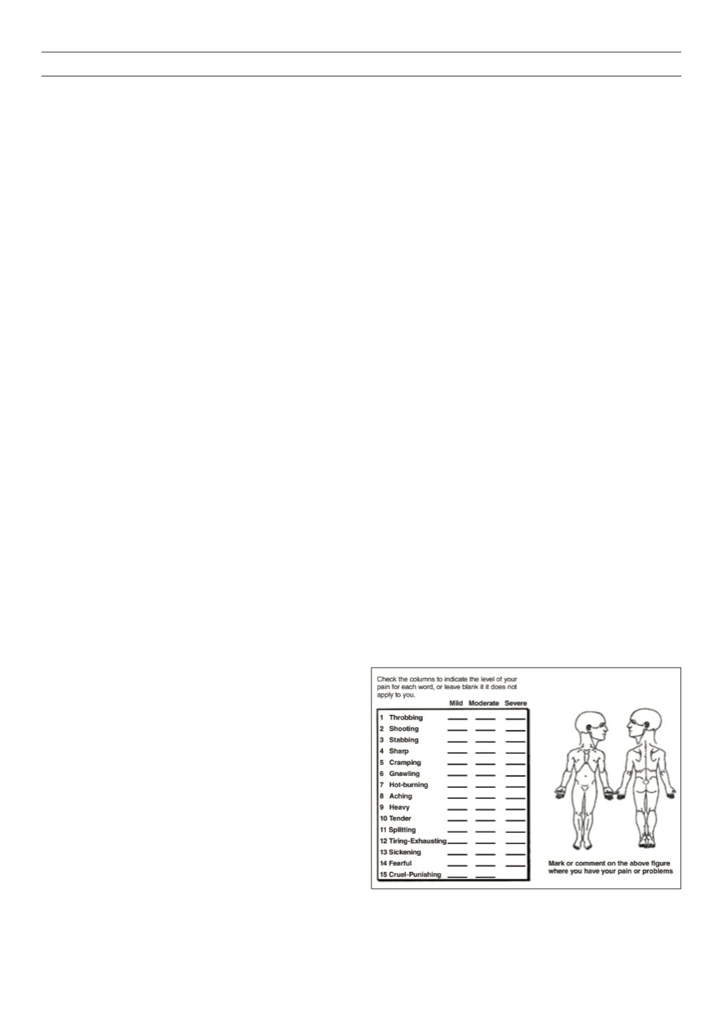
and the Brief pain inventory (BPI) (Fig. 5).
Average pain score: the Likert scale is an 11-point linear visual
analogue pain scale ranging from 0 (no pain) to 10 (maximum pain)
(Fig. 6). On examination it is imperative to assess the arterial pulses
carefully to exclude peripheral arterial disease.
Figure 5.
Severity assessment: pain diary and scales.


