REVIEW
SA JOURNAL OF DIABETES & VASCULAR DISEASE
8
VOLUME 8 NUMBER 1 • MARCH 2011
Course of vitiligo
The onset of vitiligo is insidious and asymptomatic. Many patients
become aware of depigmented areas in the spring or summer when
tanning accentuates the contrast between involved and uninvolved
skin. The course of vitiligo is unpredictable. It is usually of slow
progression, but may stabilise for a long period of time or progress
rapidly. Total body involvement that occurs within weeks or days
has been recorded. A degree of sun-induced or spontaneous
repigmentation may occur but complete, stable repigmentation is
rare.
Vitiligo and ocular disease
1,3,4
The uveal tract (iris, ciliary body and choroid) and retinal pigment
epithelium contain pigment cells. Generally vitiligo patients do not
have eye problems, but may present with uveitis.
The most severe form of uveitis occurs in Vogt-Koyanagi-Harada
syndrome, which is characterised by uveitis, aseptic meningitis, otic
involvement and vitiligo, especially of the head and neck region.
Depigmented areas involving the ocular fundus have been seen
in vitiligo, suggesting focal loss of melanocytes.
1,3,4
Although abnormal sensory hearing loss suggests impairment
of the cochlear melanocytes, there was no clear evidence of otic
abnormalities until recently. In a recent study, 37.7% of patients
were found to have hearing problems.
8
Associated disorders
1,3,4,8
Generally, people with vitiligo are healthy, but autoimmune
endocrinopathies do occur in some. The strongest association is
with thyroid dysfunction, either hyper- or hypothyroidism (Graves’
disease, Hashimoto’s thyroiditis, toxic goitre). Despite many clinical
studies, it is difficult to conclude the strength of the association
or the frequency of circulating antimicrosomal, antithyroglobulin
or anti-TSH receptor antibodies. Vitiligo may begin before, with or
after the thyroid disease, but neither the courses nor the treatments
of the thyroid disease or vitiligo seem to have any effect on each
other.
The validity of the association with diabetes mellitus has not
been established.
The incidence of Addison’s disease in vitiligo is reported to
be 2% but is likely to be less. Two of Thomas Addison’s original
patients also had vitiligo.
Pernicious anaemia is rare, but is increased in frequency in
vitiligo patients. In these patients, the vitiligo is reportedly more
widespread, and pernicious anaemia has been reported in those
with late-onset vitiligo. An increased incidence of antiparietal cell
antibodies has been documented in vitiligo patients with gastric
achlorhydria.
Depigmentation sometimes occurs in patients with malignant
melanoma, therefore sudden onset of progressive vitiligo in late
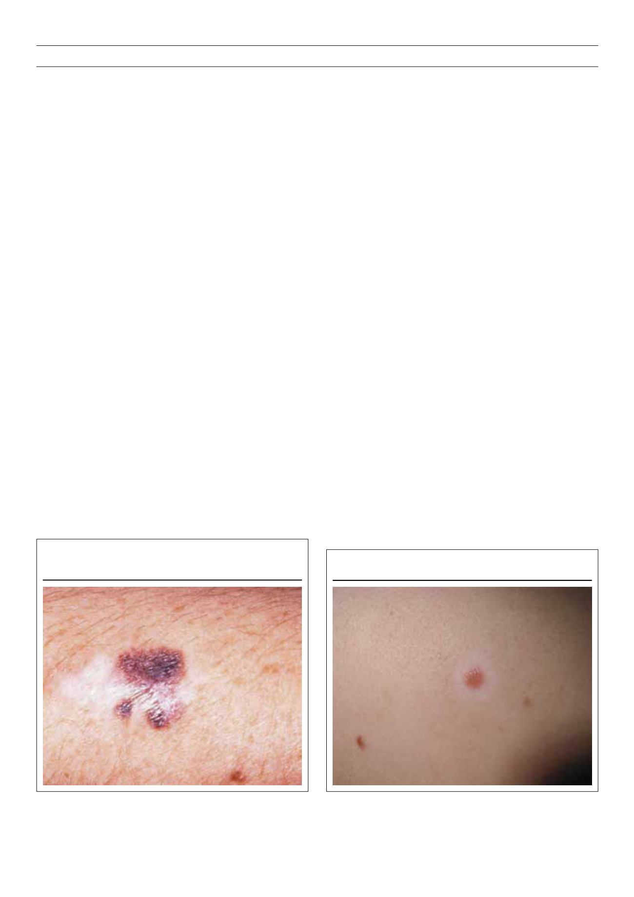
adulthood warrants a search for a suspicious lesion (Fig. 6).
9
Halo naevi (Fig. 7) occur frequently and antedate the vitiligo.
There may be one or more halo naevi and confluence of these halo
naevi leaves a vitiligo-like macule with scalloped borders.
Alopecia areata (Fig. 8) has been reported in up to 16% of vitiligo
patients and there have been reports of patients with alopecia
universalis and extensive to universal vitiligo, having associated
thyroid disease, multiple autoantibodies and hypoparathyroidism.
Autoimmune polyendocrine syndromes (APS)
Four main types of APS have been described, types 1 to 4, with
type 4 consisting of combinations not included in types 1 to 3.
1,3,4,10
The most important dermatological form is APS type 1,
which is usually due to the R257X mutation of the AIRE (auto-
immune regulator) gene on chromosome 21. It is associated
with approximately 13% of cases of Addison’s syndrome. This
condition is also called mucocutaneous candidiasis-endocrinopathy
syndrome, multiple endocrinopathy syndrome, autoimmune
polyendocrinopathy-candidiasis-ectodermal dystrophy (APECED)
syndrome or Whitaker’s syndrome. Dermatological features include:
mucocutaneous candidiasis, nail dystrophy, alopecia areata, vitiligo
and cutaneous features of Addison’s disease.
A feature in this condition is the presence of complement-fixing
melanocyte autoantibodies in almost all patients with vitiligo and
APS type 1. Autoantibodies have been described in patients with
Figure 6.
Depigmentation sometimes occurs in patients with malignant
melanoma.
Figure 7.
Halo naevus.


