REVIEW
SA JOURNAL OF DIABETES & VASCULAR DISEASE
6
VOLUME 8 NUMBER 1 • MARCH 2011
epidermis leads to oxidative degradation of catalase. Other sources
of epidermal H
2
O
2
include increased catecholamine biosynthesis
and inhibition of thioredoxin/thioredoxin reductase by calcium.
These processes may lead to melanocyte destruction.
Other theories
Other hypotheses on the aetiology of vitiligo are: reduced
melanocyte survival and dysregulation of melanocytes, destruction
of melanocytes by autocytotoxic metabolites, membrane lipid
alterations in melanocytes, deficiency of melanocyte growth
factors, destruction of melanocytes by neurochemical substances
and viral infection (e.g. cytomegalovirus).
Clinical features
Vitiligo is commonly a totally amelanotic macule, surrounded by
normal skin. The colour is uniformly milky or chalk-white. Individual
macules and patches vary in size from millimetres to centimetres.
There is an absence of inflammation or erythema (redness). The
margins of vitiligo macules are usually discrete and their borders
usually convex. Lesions may enlarge centrifugally over time and
the rate of enlargement varies from slow to rapid. Fusion of
neighbouring macules may result in complex patterns.
1,3,4,7
Variants of vitiligo
• Trichrome vitiligo: this refers to a tan zone of varying width
between the normal and totally depigmented skin. Trichrome
lesions naturally evolve to typical vitiligo macules.
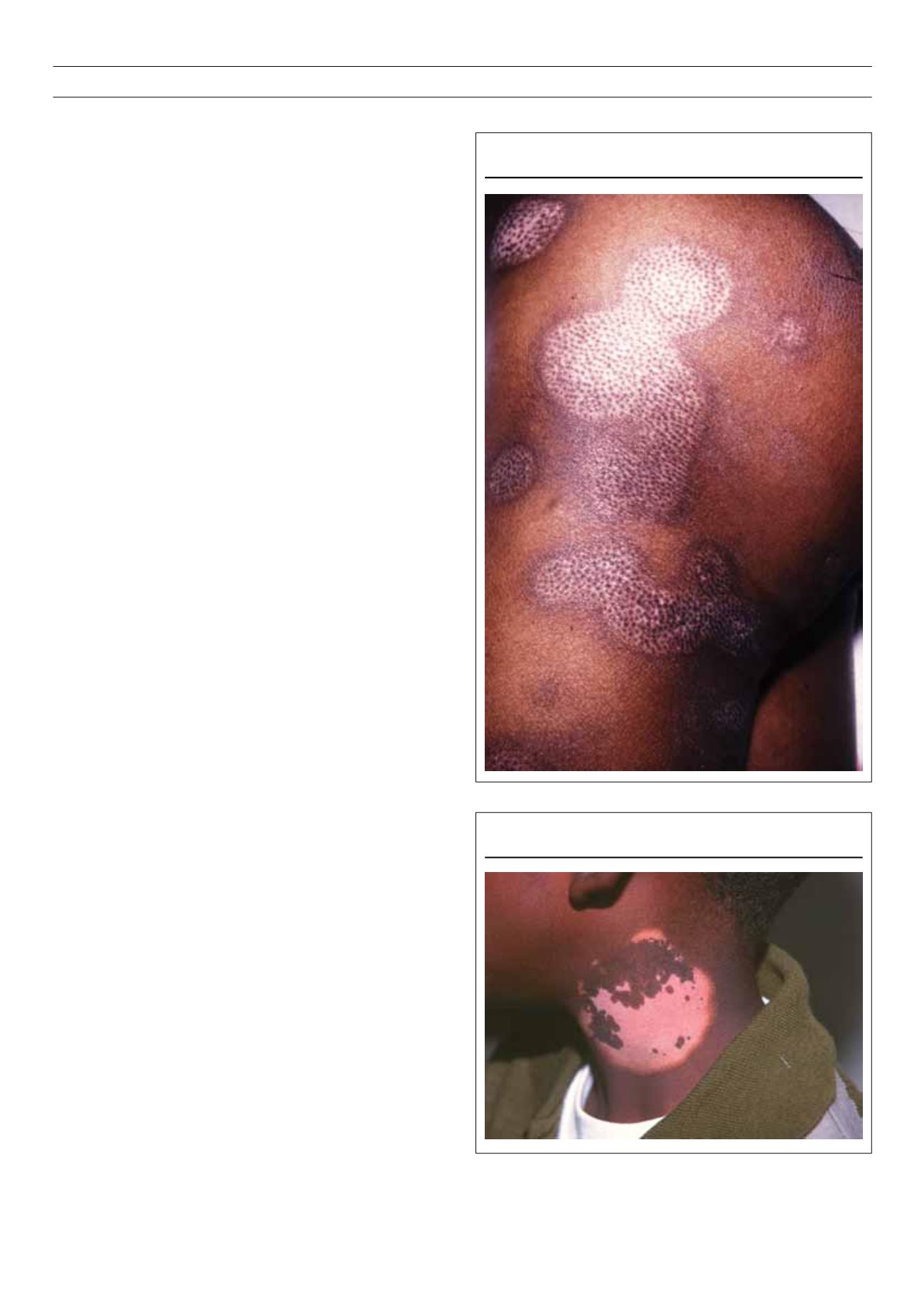
• Quadrichrome vitiligo: this refers to dark brown perifollicular
pigmentation seen in repigmenting vitiligo (Fig. 1).
• Vitiligo ponctué: this is characterised by small, confetti-like
macules.
• Inflammatory vitiligo: there is erythema of the margin of the
vitiligo macule but there is no resemblance to an inflammatory
dermatosis (Fig. 2).
Types of vitiligo
Vitiligo vulgaris/Generalised vitiligo
This is the commonest presentation. The distribution is usually
strikingly symmetrical. Although vitiligo may affect any part of the
body, characteristic patterns of involvement occur. Extensor areas,
such as the interphalangeal joints, elbows and knees are often
involved. The volar wrists, umbilicus, lumbosacral area and anterior
tibia may also be involved. These are areas subjected to repeated
trauma and friction. Areas that are normally hyperpigmented, such
as the face, axillae, groin, areolae and genitalia are also frequently
affected.
Vitiligo is sometimes peri-orificial, where it may occur around
the eyes, nose, ears, mouth and anus. Peri-ungual involvement may
be found alone, or with the involvement of certain mucosae (lips,
distal penis, nipples), called lip-tip vitiligo. Acrofacial vitiligo is the
involvement of distal digits and peri-orificial face (Fig. 3). Universal
vitiligo describes widespread involvement with few remaining
normally pigmented areas. This is the type associated with multiple
endocrinopathy syndromes.
Isomorphic Koebner phenomenon is the development of vitiligo
following trauma, such as a cut, burn or abrasion to ‘normal’
skin. The isomorphic Koebner phenomenon is more common in
progressive vitiligo and the threshold for this phenomenon appears
to be lower, such as friction from clothes.
Figure 1.
Quadrichrome vitiligo.
Figure 2.
Inflammatory vitiligo.


