SA JOURNAL OF DIABETES & VASCULAR DISEASE
REVIEW
VOLUME 8 NUMBER 1 • MARCH 2011
7
Focal vitiligo
One or more macules occur in an area, but are not clearly segmental.
Segmental vitiligo
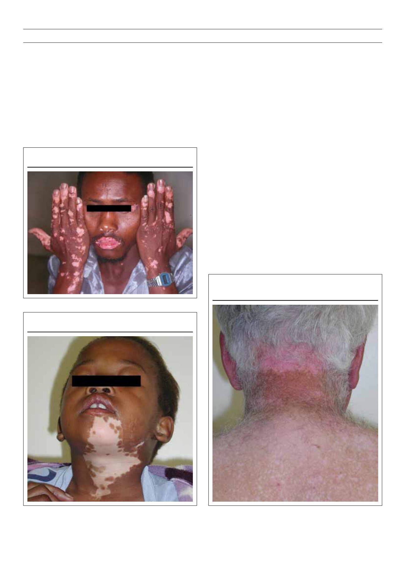
Sometimes vitiligo is unilateral and may have a dermatomal
pattern (Fig. 4). This should be considered a special type with a
stable course and is unlikely to be associated with other diseases.
Segmental vitiligo usually has an earlier onset, is more stable and
is not familial. These patients are unlikely to develop other lesions
and the isomorphic Koebner phenomenon does not occur. Five
per cent of adults and more than 20% of children have segmental
vitiligo. The trigeminal area is involved in over 50%, the neck in
23%, and trunk in 17% of patients. Up to 13% may have multiple
sites of involvement and nearly half have poliosis (white hair).
Occurrence of vitiligo
Vitiligo is less apparent in lightly pigmented people, but becomes
more obvious during Wood’s lamp examination or after tanning of
uninvolved skin (Fig. 5). Involvement of the palms and soles is less
apparent in fair-skinned individuals. In darkly pigmented persons
the contrast between vitiliginous skin and normally pigmented skin
is striking, even in habitually unexposed sites.
Spontaneous repigmentation occurs in 10 to 20% of patients,
usually in sun-exposed areas. Repigmentation occurs in younger
patients and is mainly perifollicular.
The incidence of leukotrichia (depigmented hair) varies from10%
to over 60% because of dissociated behaviour between epidermal
and follicular melanocytes. The occurrence of leukotrichia does
not correlate with the activity of the disease. Vitiligo of the scalp
may present as a localised patch of white or grey hair (usually),
total depigmentation of all scalp hair or a scattering of white hair.
Spontaneous repigmentation of depigmented hair in vitiligo does
not occur.
The commonest form of vitiligo seen in children is vitiligo vulgaris
but the frequency of segmental vitiligo is significant. The incidence
of endocrinopathies is lower than in adults, but autoantibody
formation in children with vitiligo is more significant.
Figure 4.
Segmental vitiligo.
Figure 3.
Acrofacial vitiligo.
Figure 5.
Vitiligo is less apparent in lightly pigmented people but becomes
apparent after tanning of uninvolved skin.


