REVIEW
SA JOURNAL OF DIABETES & VASCULAR DISEASE
36
VOLUME 11 NUMBER 1 • MARCH 2014
consider it as an indication for open surgical biopsy, especially if
clinical progress is unsatisfactory.
34,41,42
Culture positivity is higher
with open surgical sampling.
34,43
The diagnostic yield can be further
improved by submitting more than one specimen for culture.
44
All patients with suspected acute prosthetic joint infection (PJI)
should have a diagnostic arthrocentesis performed unless the
diagnosis is evident clinically, surgery is planned and antimicrobials
can be safely withheld before surgery. At least three and optimally
five or six periprosthetic intra-operative tissue samples or the
explanted prosthesis itself should be submitted for aerobic and
anaerobic culture at the time of surgical debridement or prosthesis
removal to maximise the chance of obtaining a microbiological
diagnosis.
45
Nucleic acid amplification tests offer potential advantages.
Molecular diagnostic methods using broad-range 16S rDNA
polymerase chain reaction (PCR) may be helpful when traditional
culture-based methods fail, especially in the context of prior
antibiotic usage. Species-specific PCR, particularly targeting
S
aureus
, can increase the sensitivity further with the additional
benefit of providing methicillin susceptibility results by amplification
of the mecA gene.
46
Studies have emphasised the difficulties associated with the
exclusion of IE solely on the basis of clinical findings. International
guidelines recommend echocardiography in
S aureus
bacteraemia
patients in order to exclude endocarditis.
40, 7
Transoesophageal
echocardiography (TOE) is often used as well as transthoracic
echocardiography (TTE) when evaluating IE as the sensitivity of TTE
is around 50%, while that for TOE approaches 100%.
48
In cases with
an initially negative TTE and TOE, repeat TTE/TOE is recommended
7–10 days later if clinical suspicion of IE remains high.
40
Imagingmodalities are also helpful in diagnosingmusculoskeletal
infection. Plain radiography is unhelpful in early joint infections but
may help exclude other causes of joint symptoms and signs. In the
case of spondylodiscitis changes may take 2–8 weeks to appear
after the onset of symptoms.
49
Early changes include subchondral
radiolucency, loss of definition of the endplate and loss of disc
height.
34,50
Later changes include destruction of the opposite
endplate, loss of vertebral height and paravertebral soft tissue mass.
In prosthetic joint infection chronic infections may cause bone loss
and evidence of loosening around an implant but these changes
are not specific to infection.
51
Computed tomography (CT) scans may yield positive findings in
the early stages. In the case of spondylodiscitis abnormalities may
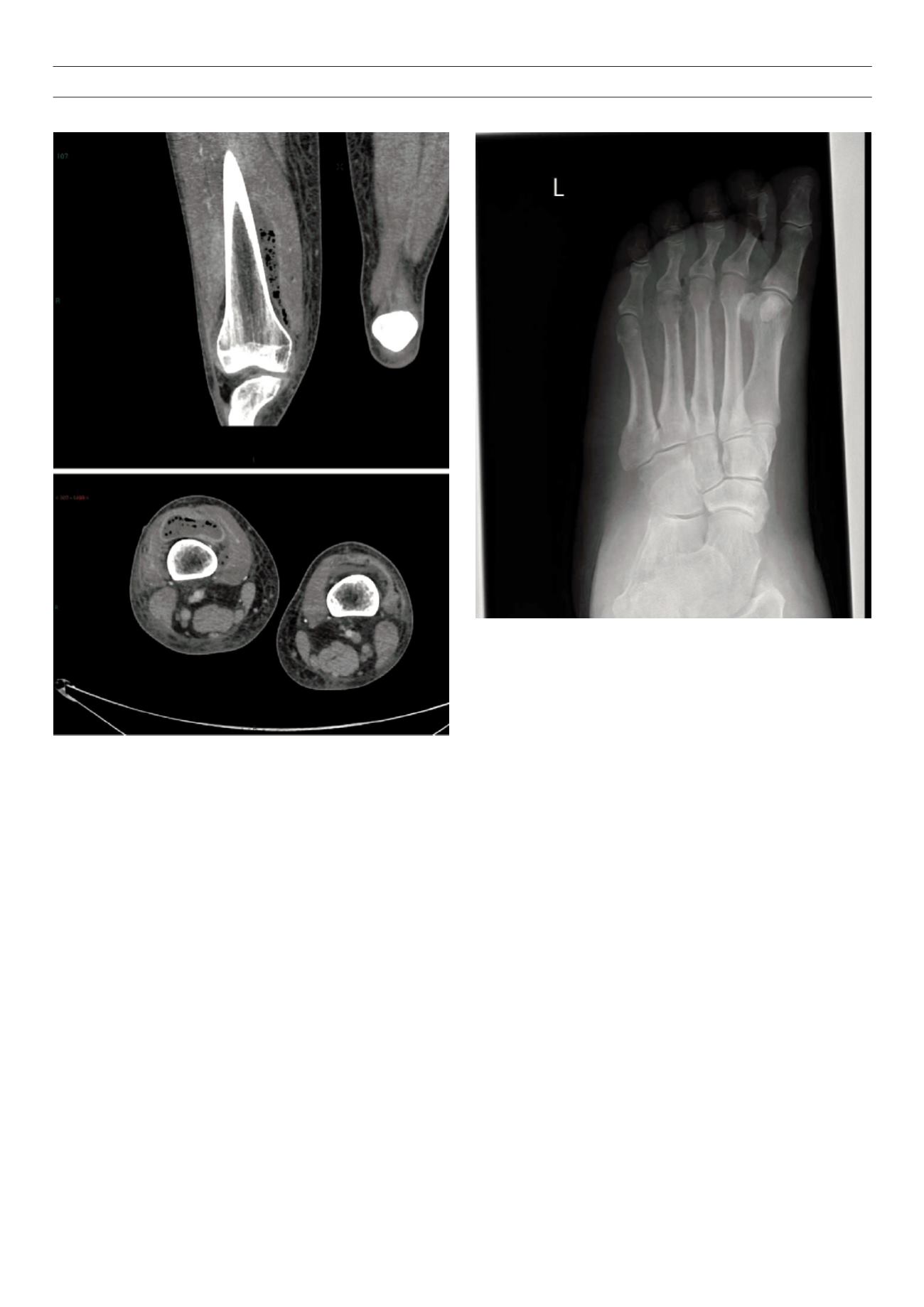
Fig. 5.
Computed tomography of thigh of a patient with type 1 diabetes who
presented with septic arthritis. Samples from arthroscopic washout isolated
S
aureus
. Despite intravenous flucloxacillin he developed worsening knee pain and
limited range of knee movement. Thigh magnetic resonance imaging showed a
large multi-loculated suprapatellar collection that extended up anteriorly as well
as along the medial aspect of the femoral shaft requiring surgical drainage.
Figure 6.
X-ray of a patient with type 1 diabetes who presented with a painful
erythematous swollen foot. A necrotic area was visible on the planter aspect
of the foot. X-ray showed gas in soft tissue around the fourth metatarsal, and
lucency is in the distal fourth metatarsal suggestive of osteomyelitis requiring
below-knee amputation for a necrotising diabetic foot infection. She had previ-
ous MRSA isolated from the foot swabs.


